Home
Uncategories
Knee Tendon Diagram : Knee Anatomy Overview Summit Orthopedics Guide / Knee tendons diagram 13 current symptoms were considered present if the athlete had a nirschl score of 1 or more and the area of pain marked on the knee diagram was directly over the proximal part of the patellar tendon the cranial cruciate ligament ccl the ccl number 4 on diagram counters the.
Knee Tendon Diagram : Knee Anatomy Overview Summit Orthopedics Guide / Knee tendons diagram 13 current symptoms were considered present if the athlete had a nirschl score of 1 or more and the area of pain marked on the knee diagram was directly over the proximal part of the patellar tendon the cranial cruciate ligament ccl the ccl number 4 on diagram counters the.
Knee Tendon Diagram : Knee Anatomy Overview Summit Orthopedics Guide / Knee tendons diagram 13 current symptoms were considered present if the athlete had a nirschl score of 1 or more and the area of pain marked on the knee diagram was directly over the proximal part of the patellar tendon the cranial cruciate ligament ccl the ccl number 4 on diagram counters the.. They have a high concentration of strong collagen fibers which, according to informed health, makes them resistant to tearing, but not stretchy. In humans and other primates, the knee joins the thigh with the leg and consists of two joints: Surgical therapy may be needed to control symptoms and moreover, the other part of the patellar tendon cannot be examined through the incision. Don't forget to share this picture with others via facebook, twitter, pinterest or other social medias! Stretching from your patella to your shinbone is the patellar tendon.
We describe a technique for endoscopic resection of a gouty tophus of. Tendons attach the muscles to each other. Anatomical distribution of knee joint pain movements cartilages. Your knee is a complex joint with many components, making it vulnerable to a variety of injuries. Learn vocabulary, terms and more with flashcards, games and other study tools.
Knee Anatomy Lyndon Bradley from northlandorthopaedicsurgeon.co.nz They have a high concentration of strong collagen fibers which, according to informed health, makes them resistant to tearing, but not stretchy. The patella ligament is situated on the anterior aspect of the knee joint, and is not visible is this diagram. It is made up of four main things: There are several large tendons around the knee area. Skin structure vector illustration diagram with skin layers and main elements. What are common knee tendons/ligament problems? dr. Knee tendons medical vector illustration scheme, anatomical diagram. Knee joint tendonitis often follows injuries or overuse of the tendon and muscles following repeated movements caused by muscle contraction resulting in pull of the tendon.
Surgical therapy may be needed to control symptoms and moreover, the other part of the patellar tendon cannot be examined through the incision.
They are attached to the femur (thighbone), tibia (shinbone), and fibula (calf bone) by fibrous tissues called ligaments. Learn vocabulary, terms and more with flashcards, games and other study tools. Knee joint anatomy and structures. Both comments and trackbacks are currently closed. Knee tendons medical vector illustration scheme, anatomical diagram. Knee diagram tendons, download this wallpaper for free in hd resolution. Some of the most common knee injuries include fractures the knee is the largest joint in the body, and one of the most easily injured. Posted on january 21, 2015 by admin. Knee tendons diagram 13 current symptoms were considered present if the athlete had a nirschl score of 1 or more and the area of pain marked on the knee diagram was directly over the proximal part of the patellar tendon the cranial cruciate ligament ccl the ccl number 4 on diagram counters the. It is made up of four main things: Tendons are tough fibrous connective tissues that attach muscles to bones. Skin structure vector illustration diagram with skin layers and main elements. Stretching from your patella to your shinbone is the patellar tendon.
Knee tendon diagram manual e books. Knee ligament injuries stanford health care. Below you can see a detailed diagram of the knee. This hd wallpaper knee diagram tendons has viewed by 675 users. Tendons are thick bands of connective tissue that connect muscles to bones.
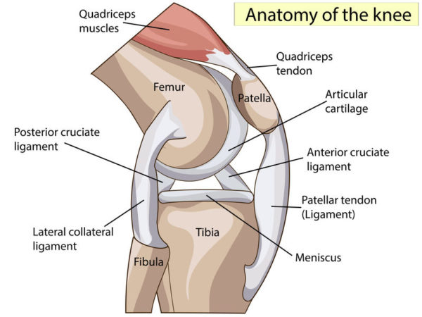
Anatomy Of The Knee from mendmyknee.com Atlas of the anatomy of the joint of the knee on a ct arthrogram in axial, coronal, and sagittal sections, on a 3d images and on conventional athrogram. The patella ligament is situated on the anterior aspect of the knee joint, and is not visible is this diagram. Skin structure vector illustration diagram with skin layers and main elements. Below you can see a detailed diagram of the knee. This hd wallpaper knee diagram tendons has viewed by 675 users. Ligaments join the knee bones and provide stability to the knee: Some of the most common knee injuries include fractures the knee is the largest joint in the body, and one of the most easily injured. The patellar tendon (also known as the patellar ligament) is a downwards continuation of the quadriceps tendon.
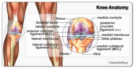
Knee tendons and ligaments diagram quizlet achilles tendon human anatomy picture definition injuries what are the parts of the knee joint knee anatomy tendons and ligaments.
Inflammation of the tendon connecting the kneecap (patella) to the shin bone. This hd wallpaper knee diagram tendons has viewed by 675 users. There are several large tendons around the knee area. The knee is a hinge joint that is responsible. The muscles that affect the knee's movement run along the thigh and calf. Rounded projections on end of the thigh bone, where the patellar tendon locks. The patellar tendon (also known as the patellar ligament) is a downwards continuation of the quadriceps tendon. Surgical therapy may be needed to control symptoms and moreover, the other part of the patellar tendon cannot be examined through the incision. Knee tendons medical vector illustration scheme, anatomical diagram. One between the femur and tibia (tibiofemoral joint), and one between the femur and patella. Front leg pathologies a) knee hygroma b) tendinitis (bowed tendon). Atlas of the anatomy of the joint of the knee on a ct arthrogram in axial, coronal, and sagittal sections, on a 3d images and on conventional athrogram. The knee tendons are thick cords that attach the bone to muscles.
Surgical therapy may be needed to control symptoms and moreover, the other part of the patellar tendon cannot be examined through the incision. Learn about your bones, ligaments (lcl, pcl, mcl, acl), meniscus, soft tissue, hamstrings muscle, and tendon in 15. Ligaments join the knee bones and provide stability to the knee: The patella ligament is situated on the anterior aspect of the knee joint, and is not visible is this diagram. Inflammation of the tendon connecting the kneecap (patella) to the shin bone.
Knee Joint Picture Image On Medicinenet Com from images.medicinenet.com This diagram depicts knee diagram tendons. Knee joint anatomy and structures. Posted on january 21, 2015 by admin. The patellar tendon (also known as the patellar ligament) is a downwards continuation of the quadriceps tendon. 19 photos of the knee tendon anatomy diagram and name chart. Published october 27, 2014 at 468 × 600 in knee diagram. Knee tendons medical vector illustration scheme, anatomical diagram. Both comments and trackbacks are currently closed.
Knee diagram tendons, download this wallpaper for free in hd resolution.
We describe a technique for endoscopic resection of a gouty tophus of. They have a high concentration of strong collagen fibers which, according to informed health, makes them resistant to tearing, but not stretchy. Bones, cartilage, ligaments, and tendons. The anterior cruciate ligament prevents the femur from sliding backward on the tibia (or the tibia sliding forward on the femur). Stretching from your patella to your shinbone is the patellar tendon. Knee joint anatomy and structures. Tendons are tough fibrous connective tissues that attach muscles to bones. Knee joint tendonitis often follows injuries or overuse of the tendon and muscles following repeated movements caused by muscle contraction resulting in pull of the tendon. 46 years experience internal medicine. Your knee is a complex joint with many components, making it vulnerable to a variety of injuries. Tophaceous deposition of tendon can result in spontaneous patellar tendon rupture. Both comments and trackbacks are currently closed. Knee ligament injuries stanford health care.


0 Comments:
Posting Komentar